missing translation for 'onlineSavingsMsg'
Learn More
Learn More
PMEL17/SILV Antibody (HMB45 + PMEL/783), Novus Biologicals™
Mouse Monoclonal Antibody
Brand: Novus Biologicals NBP2-44523-0.1MG
This item is not returnable.
View return policy
Description
Ensure accurate, reproducible results in Flow Cytometry, Immunohistochemistry (Paraffin), Immunofluorescence
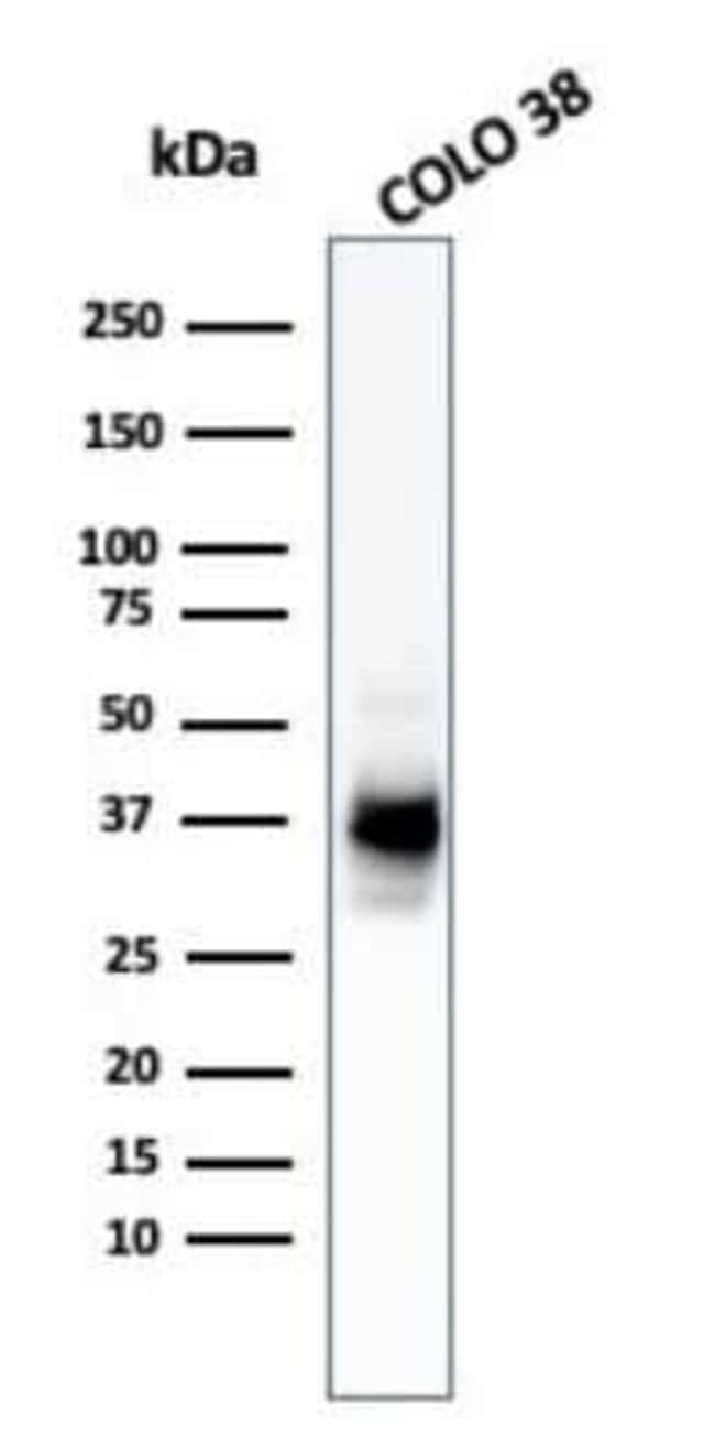
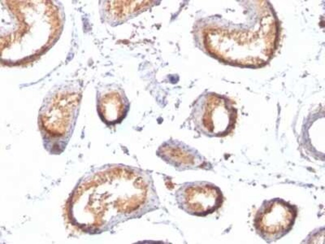
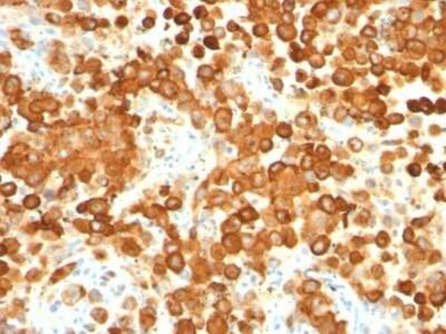
PMEL17/SILV Monoclonal specifically detects PMEL17/SILV in Human samples. It is validated for Western Blot, Immunohistochemistry, Immunohistochemistry-Paraffin.Specifications
| PMEL17/SILV | |
| Monoclonal | |
| 0.2mg/mL | |
| Flow Cytometry 0.5 - 1 ug/million cells in 0.1 ml, Immunohistochemistry-Paraffin 0.5 - 1.0 ug/ml, Immunofluorescence 0.5 - 1.0 ug/ml | |
| P40967 | |
| PMEL | |
| Extract of pigmented melanoma metastases from lymph nodes (HMB45); Recombinant human SILV protein (PMEL/783) | |
| Protein A or G purified | |
| RUO | |
| 6490 | |
| Human | |
| Purified |
| Flow Cytometry, Immunohistochemistry (Paraffin), Immunofluorescence | |
| HMB45 + PMEL/783 | |
| Unconjugated | |
| 10mM PBS and 0.05% BSA with 0.05% Sodium Azide | |
| D12S53EP1, gp100, ME20, ME20-M, melanocyte protein mel 17, Melanocyte protein Pmel 17, Melanocytes lineage-specific antigen GP100, Melanoma-associated ME20 antigen, melanosomal matrix protein17, PMEL17P100, premelanosome proteinME20M, SI, SIL, silver (mouse homolog) like, silver homolog (mouse), Silver locus protein homolog, silver, mouse, homolog of, SILVPmel17 | |
| Mouse | |
| 95 kDa | |
| 0.1 mg | |
| Primary | |
| By immunohistochemistry, it specifically recognizes a protein in melanocytes and melanomas. This MAb reacts with junctional and blue nevus cells and variably with fetal and neonatal melanocytes. Intradermal nevi, normal adult melanocytes, and non-melanocytic cells are negative. It does not stain tumor cells of epithelial, lymphoid, glial, or mesenchymal origin. Metastatic amelanotic melanoma can often be confused with a variety of poorly differentiated carcinomas, large cell lymphomas, and sarcomas using H & E stains alone. It is also difficult to differentiate melanoma from spindle cell carcinomas and various types of mesenchymal neoplasms. This MAb stains fetal and neonatal melanocytes, junctional and blue nevus cells, and malignant melanoma. This MAb also stains Angiomyolipoma (PEComa). | |
| Store at 4C. | |
| IgG |
Product Content Correction
Your input is important to us. Please complete this form to provide feedback related to the content on this product.
Product Title
For Research Use Only
Spot an opportunity for improvement?Share a Content Correction