Learn More
Description
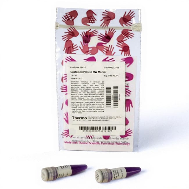
Thermo Scientific™ Unstained Protein Molecular Weight Marker is a mixture of seven native proteins (14.4kDa to 116kDa) for use as size standards in protein electrophoresis (SDS-PAGE).
Highlights:
Size range – seven proteins spanning 14.4 to 116kDa
Ready-to-use – supplied in a loading buffer for direct loading on gels
Sharp bands – equal intensity upon coomassie or silver staining
Quality tested – each lot evaluated by SDS-PAGE and Western blotting
Includes:
Proteins (0.1 to 0.2mg/mL of each) in 62.5mM Tris-HCl (pH 7.0 at 25°C), 1mM EDTA, 2% SDS, 50mM DTT, 30mM NaCl, 1mM NaN3, 0.01% bromophenol blue and 50% glycerol; Component proteins: beta-galactosidase (116kDa), bovine serum albumin (66.2kDa), ovalbumin (45.0kDa), lactate dehydrogenase (35.0kDa), REase Bsp98I (25.0kDa), beta-lactoglobulin (18.4kDa), lysozyme (14.4kDa)
Recommended for:
Accurate protein sizing on SDS-polyacrylamide gels and Western blots
Specifications
Specifications
| Content And Storage | Contents: two vials of 1 mL each Storage buffer: 62.5 mM Tris-H3PO4 (pH 7.5 at 25°C), 1mM EDTA, 2% SDS, 50 mM DTT, 30 mM NaCl, 0.01% bromophenol blue and 50% glycerol Storage: Upon receipt store at -20°C |
| Number of Markers | 7 |
| Ready to Load | Yes |
| Size Range | 14.4 to 116 kDa |
| Gel Compatibility | Bolt™ Bis-Tris Plus Gels, Novex™ Tricine Gels, Novex™ Tris-Glycine Gels, NuPAGE™ Bis-Tris Gels, NuPAGE™ Tris-Acetate Gels, SDS-PAGE Gels |
| Molecular Weight (g/mol) | 116, 66.2, 45, 35, 25, 18.4, 14.4 kDa |
| Quantity | 2 x 1 mL |
| Shipping Condition | Approved for shipment on Wet or Dry Ice |
| Product Line | Pierce |
| Product Type | Protein Ladder |
| Show More |
Frequently Asked Questions (FAQs)
The upper bands of the ladder may be degraded by proteases. Ladder, gel, buffer, pipettes, pipette tips, or equipment can be contaminated by proteases during usage. A general recommendation would be to avoid working with proteases in the same room. We would recommend preparing fresh solutions, cleaning the equipment, and using clean pipettes and tips. If the ladder itself is contaminated, please use a new tube of the ladder.
Additional bands can appear due to dithiothreitol (DTT) oxidation in the storage buffer. Please add newly prepared DTT solution to the final concentration of 100 mM and boil for 5 min at 95 degrees C. This should solve the issue. Addition of DTT is NOT recommended for prestained protein ladders, since too high a concentration of reducing agents can cause protein destaining.
No, proteins in Thermo Scientific protein ladders are not His tagged. However, non-specific interaction between the ladder proteins and primary or secondary antibodies is possible and some His-Tag detection systems, such as Thermo Scientific 6xHis Protein Tag Stain Reagent Kit, show non-specific interaction. The protein ladder bands are more readily detected when using high antibody concentrations. The non-specific cross-reactivity is difficult to predict, it often has a different pattern dependent on the antibodies used in each individual experiment. The most general way to handle this problem would be to use lower concentrations of antibodies and to use lower amount of protein ladders. It may also be useful to leave one empty well between the ladder and the sample to overcome a possible leakage of the signal to the nearby sample lane.
PageRuler Unstained protein ladders can be detected directly on Western blots by using Strep-Tactin conjugates or an antibody against the Strep-tag II sequence. All PageRuler and Spectra ladder proteins contain an integral Strep-tag II sequence, however the prestained ladders cannot be detected by Strep-Tactin conjugates.
Protein ladder bands can sometimes be detected with chemiluminescent techniques due to non-specific interactions of ladder proteins with either primary or secondary antibodies (or with both). The ladder bands are only rarely detected by chromogenic substrates. The extremely high sensitivity of the chemiluminescent assays is needed to see the bands, so the actual degree of cross-reactivity is low. The non-specific cross-reactivity is difficult to predict, it often has a different pattern depending on the antibodies used. If antibodies recognize a linear epitope, the cross-reactivity may be due to sequence homology. If antibodies react with a denaturation-resistant conformational epitope it could be impossible to identify the exact reason for detected cross-reactivity. The most general way to handle this problem would be to use lower concentrations of antibodies.
For Research Use Only. Not for use in diagnostic procedures.
By clicking Submit, you acknowledge that you may be contacted by Fisher Scientific in regards to the feedback you have provided in this form. We will not share your information for any other purposes. All contact information provided shall also be maintained in accordance with our Privacy Policy.